
Epiretinal Membrane (Macular Pucker)
Author: Albert O. Edwards, MD, PhD; Ophthalmologist and Retina Specialist
Date published: 8/5/2026
Case history: Patient with Epiretinal Left Eye
This very nice lady presented with blurring of central vision in the left eye. The right eye remained asymptomatic with normal vision. With further discussion, we found that she had each of the major symptoms patients get from epiretinal membranes. In addition to blurred vision, she had distorted vision, which she only noticed upon careful evaluation comparing the two eyes. She also had bigger images in her left eye, which we call macropsia. We further elucidated a history of her closing her left eye in order to read better because it interfered with the quality of vision out of her right eye. This is called central-peripheral rivalry. We’ll discuss these symptoms in the question-and-answer session below.
Our examination and retinal imaging confirmed an epiretinal membrane limited to the left eye. The images below compare the normal right eye with the affected left eye
Images:
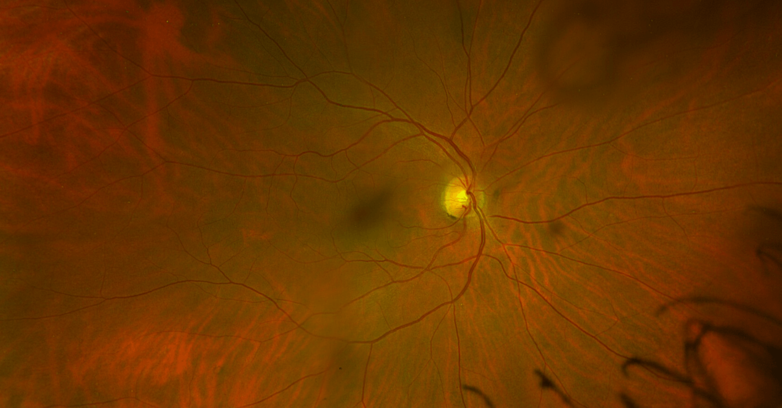
Color photograph of the inside of the back chamber of the right eye showing the optic nerve (yellow circle), retinal vessels (red vessels/lines), and the macula (central darker area). There is no epiretinal membrane in this eye. The patient has an epiretinal membrane in their other eye.
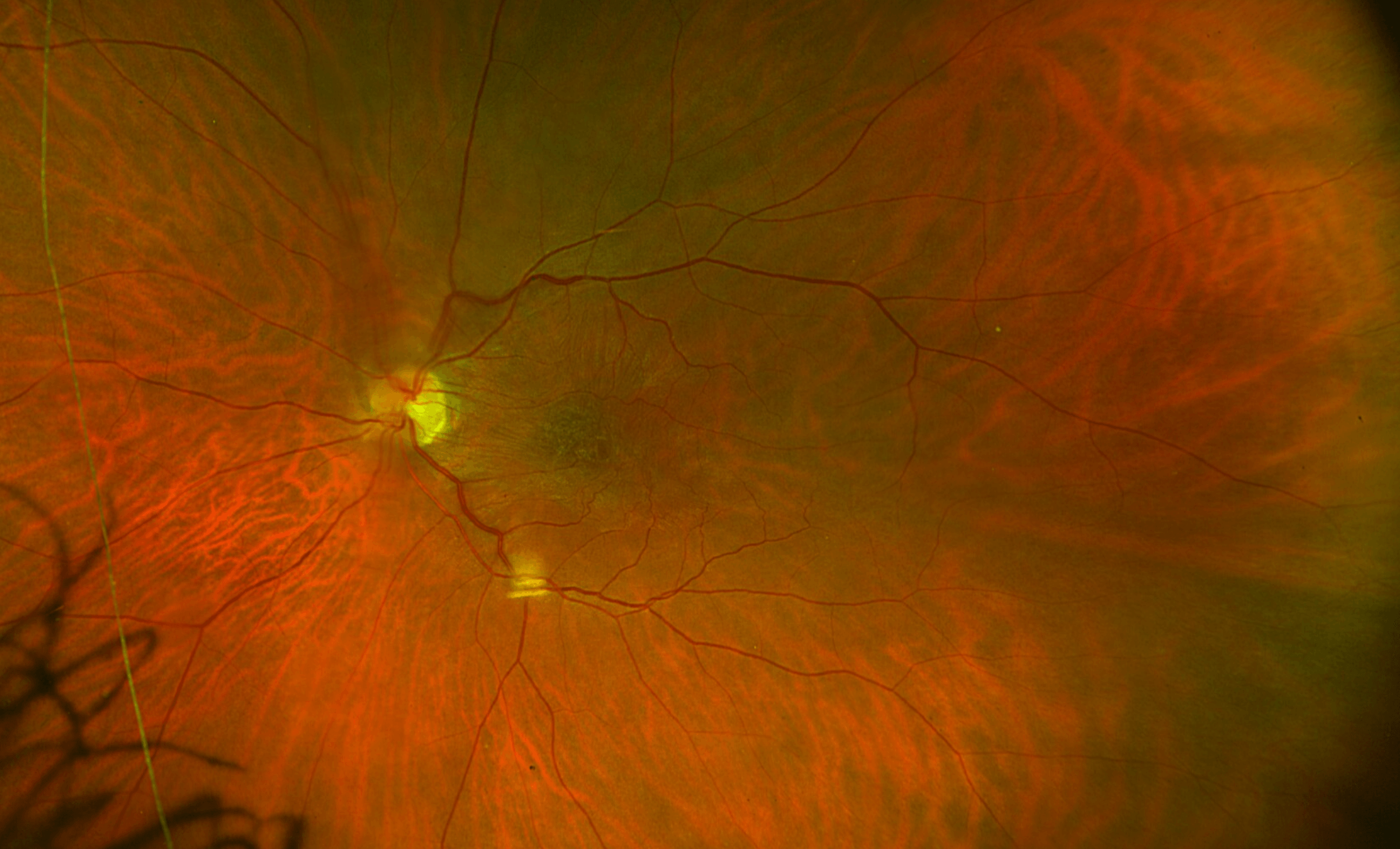
This color picture of the back of the left eye shows the wrinkling of the retina from contraction of the epiretinal membrane or macular pucker. Notice that the vessels are dragged toward the center. This leads to the patient’s symptoms. Hint: blow up the picture in your browser window to see the details better (Control +, in Windows).
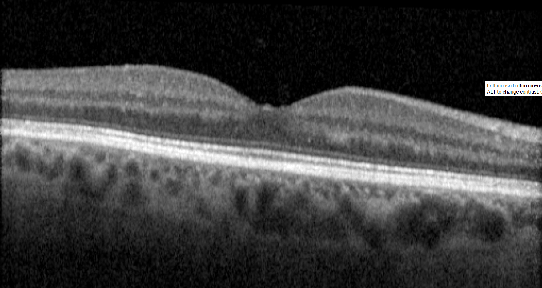
This is a cross-section through the macula of the right eye showing a normal retinal appearance. The dip in the center is the fovea, a normal finding required to have acute reading and driving vision. This patient has had a complete posterior vitreous detachment with no residual vitreous left behind on the retina that could have caused an epiretinal membrane. Compare this image to the left eye.
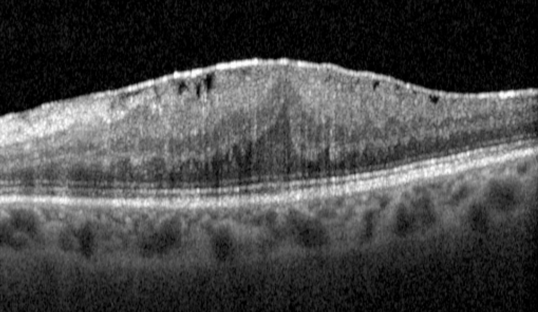
This is a cross-section through the macula of the left eye. This is an OCT (optical coherence tomogram) image. This patient had a posterior vitreous detachment, and unfortunately an outer layer of vitreous was left behind on the retina. That’s the bright white line on the top of the image. Cells infiltrated this layer of tissue. They laid down fibers that contracted, causing the folds in the retina that you see here. If you compare this left eye to the patient’s right eye, you can see that the depression is gone because it has been prolapsed toward the top of the image due to contraction of the surrounding retina. This explains the patient’s symptoms.
Frequently Asked Questions About Epiretinal Membrane
What is an epiretinal membrane? An epiretinal membrane is a thin membrane or layer of tissue that forms on the surface of the retina at the back of the eye, most often over the macula. It is also known as macular pucker or cellophane maculopathy. The membrane can contract and wrinkle the underlying retina, leading to distorted or blurred central vision. In some cases, objects may appear wavy, crooked, or bigger or closer than they actually are.
What causes an epiretinal membrane? Most epiretinal membranes develop after a posterior vitreous detachment, a common age-related change in which the vitreous gel separates from the retina. They can also form after eye surgery, ocular injury, inflammation inside the eye, retinal tears, or other retinal conditions.
What are the symptoms of an epiretinal membrane? Common symptoms include blurred vision and distorted central vision where straight lines appear wavy or bent (metamorphopsia). The contraction of the retina can lead to images appearing bigger or closer. Doctors call this macropsia. This happens because the image being projected onto the retina overlays a larger amount of retinal tissue because it has been contracted. In the other eye, which has not been contracted, the image does not project onto as large an amount of retina and thus appears smaller or further away. Peripheral vision is usually unaffected. However, some people have rivalry between the eyes because, while the peripheral retina is normally aligned in the two eyes, the central retina that we rely on to read and drive is not located in the same place in the affected eye. In this situation, images will appear to jump between the eyes, and patients will often close the eye with the epiretinal membrane in order to see better out of the other eye. One important observation is that patients present earlier with symptoms in their dominant eye. The dominant eye is the one they use to sight, for example, to take pictures with a camera. When these problems affect the non-dominant eye, the symptoms are less severe and present later with more advanced disease like in the case above.
How is an epiretinal membrane diagnosed? Diagnosis is made by the combination of the expected symptoms in conjunction with findings on examination and retinal imaging. Optical coherence tomography (OCT) provides high-resolution cross-sectional images of the retina and is the most important test for confirming the presence, thickness, and possible effect of an epiretinal membrane. Color fundus photography documents the appearance of the membrane on the retinal surface. It is important to ensure that the imaging reflects the patient’s symptoms because there are many other reasons to have blurry vision.
How is an epiretinal membrane treated? Many epiretinal membranes cause only mild symptoms and can be safely monitored with periodic examinations and OCT imaging. When vision is significantly reduced or distortion interferes with daily activities, surgery is considered. The standard procedure is a vitrectomy with careful peeling of the epiretinal membrane from the retinal surface. The internal limiting membrane, or ILM, is typically peeled in order to prevent recurrence. Chromo-vitrectomy techniques are used where the epiretinal membrane and internal limiting membrane are stained with dyes such as endocyanin green or trypan blue to make the surgery easier.
What is the outlook after epiretinal membrane surgery? Most patients experience reduced distortion and rivalry after surgery. Blurry vision will be worse initially and most patients see better than they did before surgery after several weeks. Full recovery of vision depends on the severity of the membrane and the retinal tolerance of the surgery. Can be complete recovery over months to years, but not always. Residual metamorphopsia and mild reduction in best-corrected visual acuity may persist, especially if the membrane was longstanding or caused significant retinal distortion before surgery.
Can an epiretinal membrane come back after treatment? Recurrence of an epiretinal membrane after successful surgical removal is uncommon when combined with internal limiting membrane peeling. Regular postoperative examinations help detect recurrence.
What problems can happen after surgery? Many problems can happen during or after surgery. Fortunately, most of the severe ones, like infection or massive hemorrhages, are rare. Common problems after retina surgery include:
- high or low eye pressure
- swelling or edema in the retina
- retinal tears
- retinal detachments.
Patients who have had intraocular surgery are encouraged to have regular examinations to check for these problems.
ICD10 Code: H35.37, Epiretinal membrane
CPT Code: 67042, Pars plana vitrectomy; with removal of internal limiting membrane of retina
This content is intended for patient education and is not a substitute for professional medical advice. If you are experiencing new visual distortion or blurred central vision, please schedule a retinal evaluation.