
Posterior Vitreous Detachment (PVD) and Retinal Tear following cataract surgery
Author: Albert O. Edwards, MD, PhD; Ophthalmologist and Retina Specialist
Date published: 8/7/2026
Case history: Patient with Posterior Vitreous Detachment and Retinal Tear Following Cataract Surgery
This lovely lady presented to see us several years ago for routine medical and vision eye care. She saw one of our optometrists, Dr. Fleming, who is now retired. She was referred to see me two years ago for cataract surgery due to decreasing vision in both eyes. The patient elected to have light-adjusted lenses in order to be able to have optimal distance vision in both eyes with reasonable near vision. Please see our website page at Cataract Surgery in Eugene, OR | Sterling Vision for additional information on intraocular lens options. At the time of cataract surgery, she had partial posterior vitreous detachments in both eyes as shown in Figure 1.
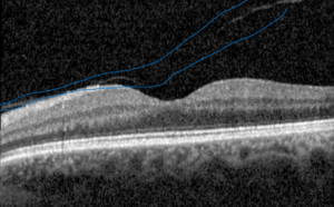
Figure 1. This optical coherence tomography (OCT) image of the left eye shows a partial posterior vitreous detachment. The vitreous gel has begun to separate from the retina in places but remains attached in others. The posterior hyaloid or surface of the gel closest to the retina has a blue circle drawn around it. This incomplete separation is a common age-related finding and was present in both of this patient’s eyes prior to cataract surgery. It is my personal belief that all patients being scheduled for cataract surgery should be evaluated with an OCT and the status and extent of vitreous detachment determined prior to surgery.
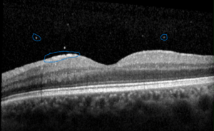
The patient was taught the importance of monitoring for the new onset of flashing lights or a significant increase in her floaters. She returned two years after cataract surgery with an acute posterior vitreous detachment, or sudden separation of the vitreous gel from the retinal surface. In addition to new-onset floaters, she had tiny, small black dots resulting from red blood cells and flashing lights from the tear and pulling on the retina from the vitreous gel. The white dots shown in Figure 2 illustrate a small amount of bleeding from the retinal tear.
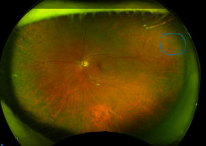
Dilated examination and color pictures of the back of the eye showed a small retinal tear in the two o’clock position. This is illustrated with a blue circle in Figure 3. This is a serious medical condition that, if not treated, can lead to a retinal detachment. A retinal detachment is where the jelly pulling on the edges of the retinal tear pumps fluid under the retina, and it blisters off like wet wallpaper. This can cause severe and permanent vision loss that cannot be corrected with glasses.
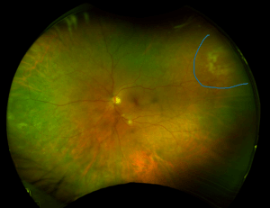
Having been taught the importance of these symptoms, the patient presented promptly to the clinic for treatment. Figure 4 shows the white burns from laser photocoagulation around the retinal tear. This will create a scar which will seal off the hole in the retina and hopefully prevent a retinal detachment. The success rate of this procedure is very high.
Frequently Asked Questions About Posterior Vitreous Detachment
What is a posterior vitreous detachment? The vitreous is the clear gel that fills the back of the eye and is attached to the retina. The vitreous gel degenerates over time and becomes filled with fluid-filled spaces and condensations of the gel, resulting in floaters. You remember seeing your floaters when you were a child, lying on your back, looking up at the sky? Well, try that now. You’ll be surprised at how many more floaters you see. With age, and more rapidly after cataract surgery, this gel can peel away from the retina. This separation is called a posterior vitreous detachment (PVD).
Why do most patients already have a partial PVD by the time they need cataract surgery? Partial vitreous separation is a common aging change. As the vitreous gel degenerates, we get floaters. Eventually, the gel collapses on itself and separates from the surface of the retina. Optical coherence tomography (OCT) imaging commonly shows incomplete separation in older adults. After cataract surgery the process often progresses more quickly. This is one of the reasons why retinal detachments are more common after cataract surgery. This is not a reason to be afraid of cataract surgery. If you need cataract surgery, you should have it. What is important here is education and access to qualified retina specialists.
How does a PVD cause a retinal tear? In some eyes, the vitreous remains abnormally adherent to the retinal surface at focal points. As the gel peels forward, these points of adhesion can tear the retina. Continued eye movement and contraction of the vitreous then tug on the tear, allowing fluid to enter beneath the retina and potentially cause a full retinal detachment. Red blood cells may also be released into the vitreous cavity from the tear, appearing as new floaters.
Why do new flashes or floaters require prompt evaluation? New-onset flashing lights or floaters can signal a retinal tear or early retinal detachment—conditions that threaten permanent vision loss if not treated quickly. Other problems can cause flashing lights and floaters. Any patient who develops these symptoms should be examined within a day or two.
Does cataract surgery increase the risk of retinal detachment? Yes. The risk of retinal detachment is higher after cataract surgery, particularly in eyes that have not already undergone a complete posterior vitreous detachment before the procedure or are very near-sighted or have high myopia. It is important for you to remember the symptoms and obtain prompt care should you develop them
Who is at the highest risk of retinal detachment from a PVD? Patients with high myopia (severe nearsightedness) face a substantially elevated risk compared with those who are not highly myopic. These individuals need especially careful education about symptoms and closer follow-up. Retinal tears appear to be present in about 3 out of 100 people, based on autopsy studies. The rate of retinal detachment in the population is much lower, approximately one in 1,000 people. Even though the odds of having a retinal detachment after a posterior vitreous detachment are low, failure to prevent one will lead to unpleasant and complicated surgery and the potential for permanent vision loss. Retinal detachments are a very serious medical condition.
How is a retinal tear from PVD treated? When a retinal tear is found, prompt in-office laser photocoagulation is used to create a barrier of scars around the tear. This seals the retina and prevents fluid from entering underneath it, greatly reducing the chance of progression to a retinal detachment.
What is the outlook after laser treatment of a retinal tear? Most patients do very well after successful laser treatment and do not develop a retinal detachment. Close follow-up is important. A residual layer of vitreous left on the retinal surface after the detachment may occasionally lead to an epiretinal membrane over time, which is why ongoing monitoring is recommended.
Should you know the stage of your PVD? I believe the answer is yes. There is a four-step staging system ranging from no PVD to early partial PVD to a nearly complete partial PVD to a complete PVD. These are the steps through which we go from not having a posterior vitreous attachment to having one. Knowing where we are along this step gives us some idea of our risk of developing one over the next few years. There’s no good way to know exactly how long that will take, but knowing that you’re at risk is important and you can ensure that you have access to appropriate care in a timely manner
ICD10 Codes:
H43.81, Vitreous degeneration / posterior vitreous detachment
H33.3-, Retinal breaks without detachment
CPT Code: 67145, Prophylaxis of retinal detachment without drainage; photocoagulation
This content is intended for patient education and is not a substitute for professional medical advice. If you are experiencing new flashing lights or floaters, please contact your eye-care provider promptly for evaluation.