
Diabetic Macular Edema (DME)
Reviewed and written by Albert O. Edwards, MD, PhD, Retina Specialist and Ophthalmologist
Last reviewed: October 2026
In short: Diabetic macular edema (DME) is swelling of the macula, the center of the retina, caused by fluid leaking from blood vessels damaged by diabetes. It can blur central vision, and sometimes be present without any symptoms. A comprehensive eye exam and an OCT scan can find it, and treatment can often reduce the swelling and protect your vision.
Diabetic macular edema (DME) is swelling in the macula, the small central part of the retina that you use to read, drive, and recognize faces. It happens when blood vessels damaged by diabetes leak fluid into the retina. DME is a complication of diabetic retinopathy, and finding it early gives you more options to protect your vision.
Watch: Dr. Edwards explains diabetic macular edema
Video: Dr. Albert O. Edwards, MD, PhD, Retina Specialist, explains diabetic macular edema (4:44). English captions are available in the player. Watch on YouTube
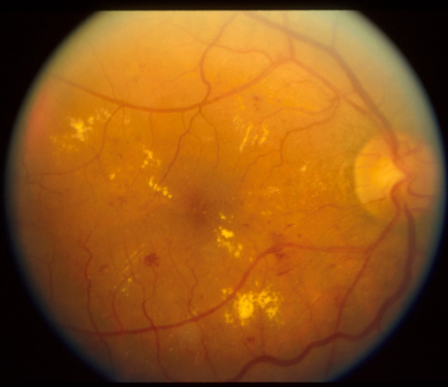 Figure 1. This is a color fundus photograph of the macula in a patient’s right eye. The photograph is taken through a dilated pupil and shows the inner surface of the back of the eyeball. The disc-shaped yellow structure on the right is the optic nerve. The reddish lines are retinal arteries and veins. The center of the picture, where it’s darker, shows the fovea, and most of the region shown is the macula. The yellowish condensations are aggregations of protein and fat from blood fluid that leaked out of capillaries damaged by diabetes. As the blood fluid that causes DME is reabsorbed, the fat and protein are left behind. You can also see reddish spots and blotches that are microaneurysms and hemorrhages. Microaneurysms are circular dilations of capillaries, the smallest level of blood vessels in the body. These are very common in diabetes. Image courtesy of Sterling Vision
How DME Develops
Over time, high blood sugar can damage the tiny blood vessels (called capillaries) in the retina. The retina is the light-sensitive tissue at the back of the eye. Damaged vessels can leak fluid. When fluid builds up in the macula, the macula swells and central vision can become blurry. Accumulation of fluid in the macula is called DME.
Symptoms
- Blurry or wavy vision in the center of what you see
- Colors that look faded or washed out
- Objects that look a different size with each eye
Some people have no symptoms at first. A comprehensive eye exam can find changes before you notice them.
Risk Factors
- Anyone with diabetes, including type 1, type 2, and gestational diabetes
- People who have had diabetes for a long time
- People with high blood sugar, high blood pressure, or metabolic abnormalities such as high cholesterol
- People with diabetes who are pregnant, or who are pregnant and develop diabetes
Diagnosis
- Comprehensive eye exam: your doctor examines the retina, and may use eye drops to widen your pupils.
- Optical coherence tomography (OCT): a quick, painless scan that shows the layers of the retina and whether the macula is swollen.
- Color photographs: photographs may be taken of the retina to allow careful inspection for microaneurysms, hemorrhages, and other more advanced vascular damage from diabetes.
- Fluorescein angiogram: a dye is placed in a vein in your arm, and photos show where blood vessels in the retina are leaking.
- Amsler grid: a simple grid of lines that can help you notice changes in your central vision. This vision test is typically used by patients at home.
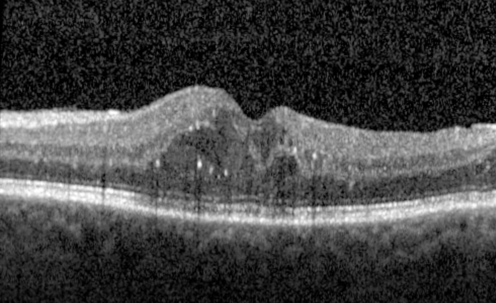 Figure 2. The image shows a slice through the retina created using light. This is called an optical coherence tomogram or OCT. This scan is from a Sterling Vision patient with diabetic macular edema, taken before treatment. The clear dark spaces in the retina are cystic collections of fluid. The fluid makes the retina thicker and raises the center of the macula, the fovea, which is normally a small dip. When these fluid pockets are seen in diabetic patients, they are called diabetic macular edema, or DME. Image courtesy of Sterling Vision
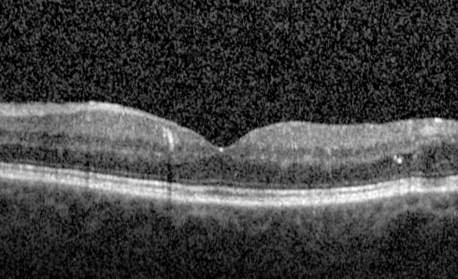 Figure 3. This OCT slice is from the same eye after treatment with anti-VEGF injections twice at 4-week intervals. The dark fluid spaces seen in Figure 2 are no longer visible, the retina is thinner, and the normal dip at the center of the macula, the fovea, has returned. Note: VEGF = Vascular Endothelial Growth Factor. Image courtesy of Sterling Vision
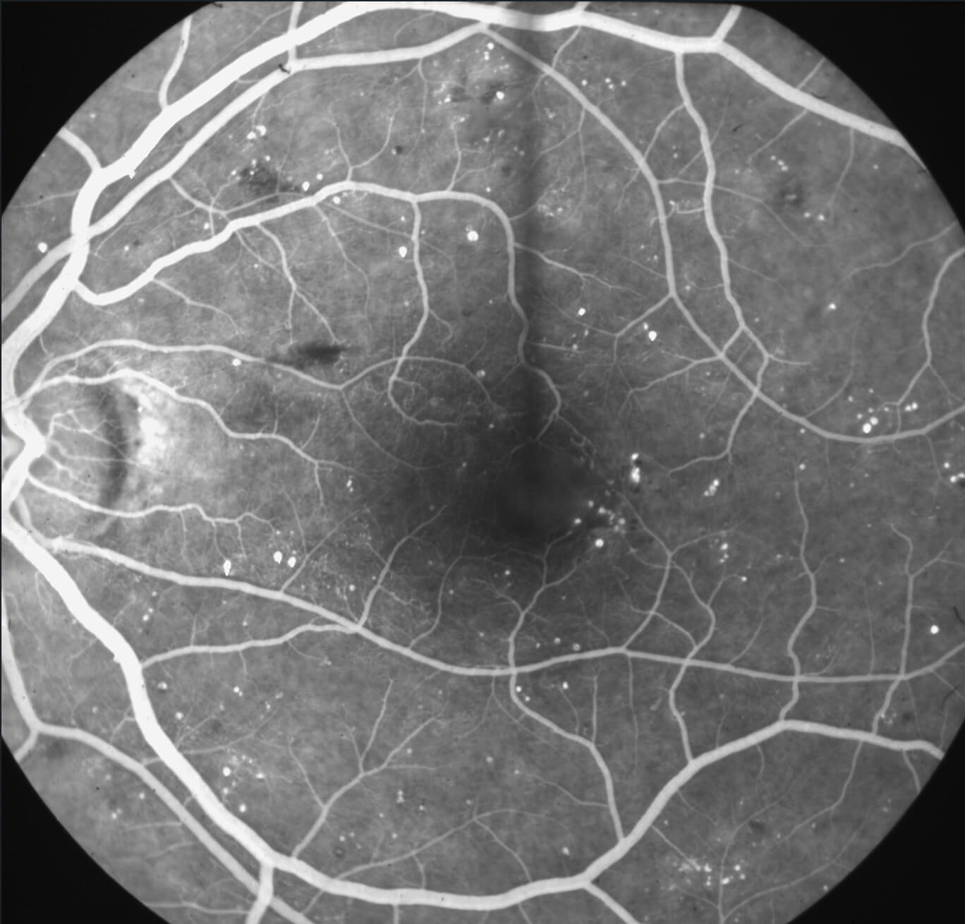 Figure 4. This is a mid-phase fluorescein angiogram of the left eye of a patient with diabetic retinopathy with minimal DME. The round, circular, bright white structures are microaneurysms that have filled with the fluorescein dye that was injected into this patient’s arm vein approximately 2 minutes before this picture was taken. You can see the very fine capillaries around the very center of the image are irregular in shape, and some of them are missing. This is called capillary dropout, where the capillaries are lost. In the upper right-hand corner, you can see a ground-glass-appearing area with minimal microaneurysms or hemorrhages. The capillaries have been lost here, which is why no retinopathy is seen. The dark streaks are flame hemorrhages. The dark blots are blot hemorrhages. This patient has significant diabetic retinopathy and needs to be followed multiple times a year for the development of problems that may be irreversible if not treated in a timely manner. The patient does not need treatment for DME at this time but is at high risk for developing DME. Image courtesy of Sterling Vision
Treatment Options
Your Retina Specialist will talk with you about treatment options. While there are multiple options, there’s likely only one that is a good fit in a particular situation:
- Anti-VEGF injections: medicine placed in the eye with a very fine needle after the eye is numbed. These medicines can reduce swelling and are usually given as a series of visits over time.
- Steroid medicines: given as an injection or a small implant in the eye to reduce swelling.
- Laser treatment: used in some cases to help reduce leaking.
- Vitrectomy surgery: used in some cases.
Working with your primary care doctor to manage blood sugar, blood pressure, diet, and improve your metabolic state, including lipid levels, also helps protect your eyes.
Clinical Research
Some patients may be eligible to participate in clinical research studies through our affiliated research program, Verum Research. Ask your Retina Specialist whether learning about research is right for you. Taking part is always your choice, and your regular care does not depend on it. You can learn about clinical trials at the government website called clinicaltrials.gov.
Role of the Retina Specialist
If you have diabetes and notice blurry or wavy central vision, or if your optometrist or another eye doctor has found swelling in your macula, schedule a visit with one of our Retina Specialists. The Sterling Vision Retina Care Team works at our Valley River Plaza (Eugene) and Thurston (Springfield) offices.
Frequently Asked Questions
Can I have DME without noticing symptoms?
Yes. Diabetic eye disease can be present before you notice any change in your vision. That is why people with diabetes are advised to have a comprehensive eye exam at least once a year, or as often as their eye doctor recommends.
Should I be afraid of eye injections?
No. Delaying or stopping treatment can lead to vision loss, and some of that loss may not come back even after treatment restarts. Fear of injections is one reason some patients miss treatment. Our Retina Care Team has developed a sophisticated protocol for ensuring your comfort during and after the injections. Injections use very small and thin needles a bit thicker than a hair, and the eye is carefully numbed in order to prevent discomfort.
How well do the injections work?
The anti-VEGF injections are miracle drugs. They have extremely low side effects. The eye tolerates them very well. They will completely suppress some of the manifestations of diabetic eye disease and will markedly reduce swelling and often will improve vision. Since we began using these drugs for diabetic eye disease in 2005, patients have been dramatically better off. Of course, not all patients respond well to any treatment, and this is the role of Retina Specialists with advanced training in diabetic eye disease.
How often should I have my eyes checked if I have diabetes?
Get a diabetic eye exam at least once a year, or more often if your eye doctor recommends it. If you are pregnant and have newly diagnosed diabetes, get an eye exam as soon as possible. If you already have DME or diabetic retinopathy, our Retina Care Team will set a schedule for you.
What can I do to reduce my chance of getting DME?
The most important steps are keeping your blood sugar (A1C), blood pressure, and blood lipids/cholesterol in the range your doctor recommends. Of course, you should not smoke. Regular physical activity and a healthy diet help with all three. Just as important is a comprehensive eye exam at least once a year, so advanced disease can be found and treated before it affects your vision. Remember, a great deal of damage can be done to the eye without a patient having any symptoms. The first symptom could be the beginning of a long and complicated process involving eye injections and retina surgery to try to save some of your vision–all of which could have been avoided by regular eye examinations and earlier intervention.
Is DME reversible, or will it come back?
Treatment can often reduce or clear the swelling, and many patients keep or even improve their vision. Vision that has already been lost from long-standing damage may not fully return. DME is a chronic condition, so the swelling can come back, which is why regular follow-up visits matter even when your eyes feel fine. In addition to improving DME, the anti-VEGF injections reduce the severity of the diabetic retinopathy. The need for injections typically goes down over time. In large clinical trials, the number of injections needed usually fell after the first year, and many patients needed fewer injections in later years. Even when fewer injections are needed, regular follow-up visits remain important. Long-term control of blood glucose levels and improving metabolic health are very important for reducing diabetic eye disease and the need for treatment. Please speak with your endocrinologist, your primary diabetic care doctor or practitioner.
Can DME make me go blind?
Without treatment, DME can cause severe loss of central vision, the vision you use for reading, driving and recognizing faces. With early detection and timely treatment, the risk of serious vision loss is much lower.
Will I need injections forever?
Not always. Most patients start with injections about once a month. As the swelling improves, many can space them out, and some can eventually stop. Others need ongoing treatment to protect their vision. Your Retina Specialist will tailor the schedule to how your eyes respond.
How soon will my vision improve after treatment?
Improvement is usually gradual. Injections do not change vision right away, and it can take several months of treatment for the swelling to go down. Keeping every scheduled visit gives the treatment the best chance to work.
Can DME go away on its own?
Mild swelling away from the center of the macula sometimes stays stable, and your Retina Specialist may recommend watching it closely rather than treating it right away. Swelling that involves the center of vision usually needs treatment. Either way, close monitoring is important because DME can change between visits. Improving your blood glucose levels, metabolic health, diet, and addressing any fluid retention in your body can reduce DME.
What is normal after an injection, and when should I call?
Mild irritation, a scratchy feeling, a red spot on the white of the eye and small floating bubbles or spots are common and usually clear within a day or a few days. The vision is always worse right after an eye injection, but it should improve slowly over hours or a day or two. The most feared complication of an eye injection occurs in about 1 in 5,000 injections. It is called endophthalmitis and is a bacterial infection inside of the eye. It typically starts about 3 to 14 days after the injection. Infection does not show up on the day of the injection or the next day. If your eye is red or uncomfortable the day after an injection, that is most likely inflammation, not infection; you still need to come see us for an urgent appointment and treatment. Infection or inflammation presents with decreased vision that progresses very rapidly. That means the vision is getting worse hour by hour. If it started overnight, you might wake up with severe vision loss. It is often painless but may be associated with sensitivity to light. We’re not talking about a few floaters or a slight worsening in your floaters, but rather an impressive and rapidly progressing loss of vision. Call us right away at 541-342-2201 to arrange a time to come into the office if you have increasing pain, increasing redness, light sensitivity or progressively worsening vision after an injection, because these can be signs of infection. A member of the Sterling Vision Retina Care Team is highly skilled in the management of complications of diabetic retinopathy and eye injections, and one of us is almost always available to assist with your urgent eye care needs at our Valley River Plaza office. Please let us know you’re having a problem, and we’ll get you into the office for treatment.
Can I drive or go back to work after an injection?
Most people can return to their normal activities, including work, the same day. Because your pupils may be dilated and your vision may be blurry for a few hours, plan to have someone drive you home from your injection visit until you know you won’t need help.
Will my insurance cover treatment?
Treatment for DME is a medical service, and most insurance plans, including Medicare, cover it. Some plans, especially Medicare Advantage plans, require prior authorization before injections. Our team will check your coverage before treatment. To book an exam, call us at 541-342-2201.
How is DME different from macular degeneration?
Both affect the macula, the part of the retina responsible for sharp central vision, but they have different causes. DME is swelling caused by leaking blood vessels damaged by diabetes. Age-related macular degeneration (AMD) is an age-related disease, and in its wet form abnormal new blood vessels grow under the retina and leak. Some of the same injection medicines are used for both.
If I control my blood sugar, will the swelling go away?
Good control of blood sugar, blood pressure and cholesterol helps protect your eyes and lowers the chance of DME getting worse, but it usually does not make existing swelling go away by itself. Most patients need both good diabetes and metabolic control and eye treatment. Please work diligently with your diabetic doctor or practitioner.
Does DME affect both eyes?
DME can affect one or both eyes. When only one eye is affected, the other eye can hide the problem, so changes may go unnoticed for a long time. Checking each eye separately, by covering one eye at a time, and having regular comprehensive eye exams help catch it early. The Amsler grid mentioned above is a tool that can be used at home to check your vision.
Can I have cataract surgery if I have DME?
Yes. Many patients with DME have successful cataract surgery. Because surgery can sometimes make macular swelling worse, your Retina Specialist and cataract surgeon will usually want the swelling under control first and may treat it before or around the time of surgery.
Learn more about our Retina Care Team at Sterling Vision.
Book AppointmentCall 541-342-2201
Sources
This page is general patient education based on information from these organizations. It is not a substitute for an exam. Your doctor will talk with you about your own eyes. The primary goal of this education is to reduce vision loss in patients with diabetes by reducing fear of treatment and increasing compliance with established recommendations for diabetic eye exams.
- The Sterling Vision Retina Care Team
- National Eye Institute: Macular Edema
- National Eye Institute: Diabetic Retinopathy
- American Academy of Ophthalmology: What Is Macular Edema?
- American Academy of Ophthalmology: Diabetic Eye Disease
- Lim JI, Kim SJ, Bailey ST, et al. Diabetic Retinopathy Preferred Practice Pattern®. American Academy of Ophthalmology. Ophthalmology. 2025;132(4):P75-P162 (PubMed)
- Browning DJ, Stewart MW, Lee C. Diabetic macular edema: evidence-based management. Indian J Ophthalmol. 2018;66(12):1736-1750 (PubMed)
- Chauhan MZ, Rather PA, Samarah SM, Elhusseiny AM, Sallam AB. Current and novel therapeutic approaches for treatment of diabetic macular edema. Cells. 2022;11(12):1950 (PubMed)
- American Academy of Ophthalmology: What Is Optical Coherence Tomography?
- American Academy of Ophthalmology: What Is Fluorescein Angiography?
- Tripathy K, Salini B. Amsler Grid. In: StatPearls. Treasure Island (FL): StatPearls Publishing; 2023 (PubMed)
- Elman MJ, Ayala A, Bressler NM, et al; Diabetic Retinopathy Clinical Research Network. Intravitreal ranibizumab for diabetic macular edema with prompt versus deferred laser treatment: 5-year randomized trial results. Ophthalmology. 2015;122(2):375-81 (PubMed)
- Diabetic Retinopathy Clinical Research Network; Elman MJ, Qin H, Aiello LP, et al. Intravitreal ranibizumab for diabetic macular edema with prompt versus deferred laser treatment: three-year randomized trial results. Ophthalmology. 2012;119(11):2312-8 (PubMed)
- Schmidt-Erfurth U, Lang GE, Holz FG, et al; RESTORE Extension Study Group. Three-year outcomes of individualized ranibizumab treatment in patients with diabetic macular edema: the RESTORE extension study. Ophthalmology. 2014;121(5):1045-53 (PubMed)
- Zhou C, Shen Y, Li S, et al. Persistent vision loss of eyes lost to follow-up with diabetic macular edema receiving intravitreal anti-VEGF therapy. Ophthalmol Sci. 2026;6(4):101107 (PubMed)
- Shahzad H, Mahmood S, McGee S, et al. Non-adherence and non-persistence to intravitreal anti-vascular endothelial growth factor (anti-VEGF) therapy: a systematic review and meta-analysis. Syst Rev. 2023;12(1):92 (PubMed)
- National Eye Institute: Injections to Treat Eye Conditions
Further Reading
- American Society of Retina Specialists: Macular Edema (patient handout, PDF)
- American Society of Retina Specialists: Diabetic Retinopathy
- EyeWiki (American Academy of Ophthalmology): Diabetic Macular Edema
- EyeWiki (American Academy of Ophthalmology): Diabetic Retinopathy
- PubMed: Research articles on diabetic macular edema
- ClinicalTrials.gov: Clinical trials for diabetic macular edema