
Non-Proliferative Diabetic Retinopathy (NPDR)
Reviewed and written by Albert O. Edwards, MD, PhD, Retina Specialist and Ophthalmologist
Last reviewed: October 2026
In short: Non-proliferative diabetic retinopathy (NPDR) is the earlier stages of diabetic eye disease, when diabetes damages the small blood vessels in the retina. It typically causes no symptoms. A comprehensive eye exam can find it, and the severity tells your doctor your risk of vision-threatening problems and how often you need checkups.
Diabetic retinopathy is an eye disease that can affect anyone with diabetes. It damages the small blood vessels in the retina called capillaries, the light-sensitive tissue at the back of the eye (1). People with diabetic retinopathy first have the early stages, called non-proliferative diabetic retinopathy (NPDR), and it often causes no symptoms (2). Regular comprehensive eye exams help find it early (3). This is important because the severity of NPDR determines the risk of vision-threatening complications and how frequently you should have eye examinations to prevent permanent damage to your vision and eye (4, 5). Just because NPDR is asymptomatic or “early” does not mean that knowing its severity is not important.
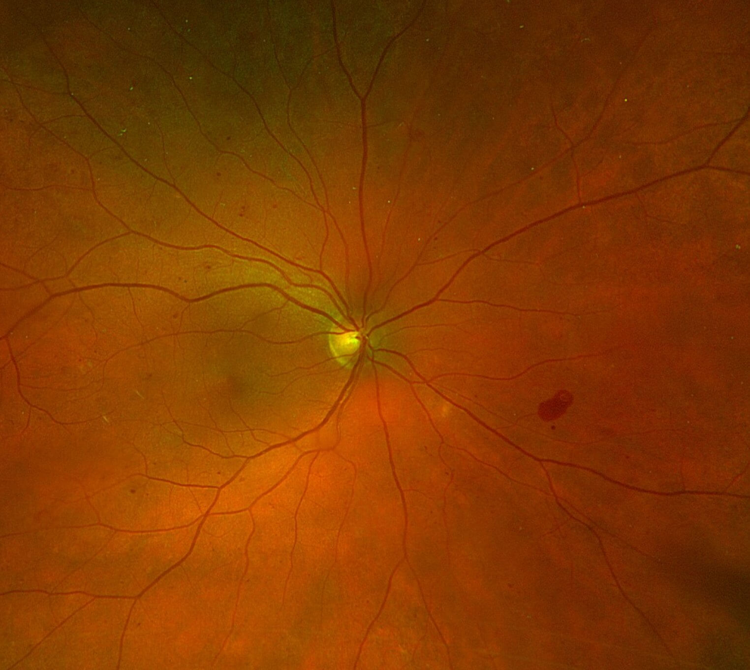
Figure 1. This is a color fundus photograph of the right eye of a patient with non-proliferative diabetic retinopathy. The yellow circle is the optic disc. The red-colored lines coming off the disc are the retinal arteries, which are smaller and lighter-colored, and the retinal veins, which are darker and larger. The arteries take blood into the eye, and the veins drain it out of the eye. The tiny red dots scattered across the retina are microaneurysms and small hemorrhages. The red blotch on the right-hand side of the image is a large blot hemorrhage from a broken capillary. Image courtesy of Sterling Vision.
Stages of Diabetic Retinopathy
Diabetic retinopathy has two main stages:
- Non-proliferative diabetic retinopathy (NPDR): the early stage. Blood vessel walls in the retina weaken. Tiny bulges can leak fluid and blood, and some vessels can close off (2, 6). Fluid can build up in the macula, the center of the retina, causing diabetic macular edema (DME).
- Proliferative diabetic retinopathy (PDR): the advanced stage. New, abnormal blood vessels grow on the retina. They can bleed into the gel inside the eye, form scar tissue, and in some cases lead to retinal detachment or a severe form of glaucoma called neovascular angle-closure glaucoma (7).
Not everyone with NPDR goes on to the advanced stage (4). Regular exams let your doctor determine the severity of NPDR (8). I call this your “DR score.” Just like you know your Hemoglobin A1C level: you should know your “DR score”.
Symptoms
Early NPDR usually has no symptoms. As the disease gets worse, you may notice:
The importance of knowing your “DR score” is to ensure that you get the care you need to prevent permanent loss of vision and avoid unpleasant and complicated vitreo-retinal surgical procedures if diabetic retinopathy is not properly managed.
Risk Factors
- Anyone with diabetes, including type 1, type 2, and gestational diabetes (6)
- The risk goes up the longer you have diabetes (9)
- High blood sugar, high blood pressure, and metabolic dysfunction such as high cholesterol raise the risk (7)
- People with diabetes who become pregnant should get an eye exam as soon as possible (10)
Diagnosis
- Comprehensive eye exam: your doctor looks at the blood vessels in the retina. The doctor will also look for blood vessels in the front of your eye, on the iris, and possibly in the angle of your eye if your eye pressure is elevated (11).
- Color pictures of the retina: color pictures are excellent screening tools for looking at the retina carefully to determine the “DR score.” (12, 13)
- OCT: images that show the retina cross section, including whether the macula is swollen (14).
- Fluorescein angiogram: a dye test that shows blood vessels that are leaking or blocked (15).
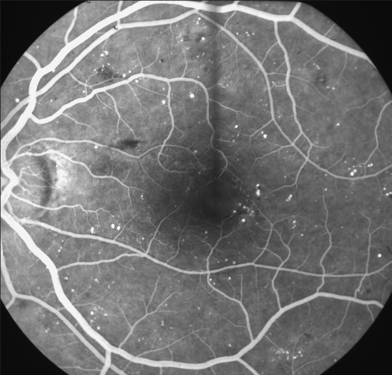
Figure 2. This is a mid-phase fluorescein angiogram of the left eye of a patient with diabetic retinopathy. The round, circular, bright white structures are microaneurysms that have filled with the fluorescein dye that was injected into this patient’s arm vein approximately 2 minutes before this picture was taken. You can see the very fine capillaries around the very center of the image are irregular in shape, and some of them are missing. This is called capillary dropout, where the capillaries are lost. In the upper right-hand corner, you can see a ground-glass-appearing area with minimal microaneurysms or hemorrhages. The capillaries have been lost here, which is why no retinopathy is seen. The dark streaks are flame hemorrhages. The dark blots are blot hemorrhages, which are seen in diabetes. This patient has significant diabetic retinopathy and needs to be followed multiple times a year for the development of problems that may be irreversible if not treated in a timely manner. Image courtesy of Sterling Vision.
Treatment Options
For early NPDR, the usual plan is monitoring with regular exams, along with managing blood sugar, blood pressure, and metabolic dysfunction including cholesterol with your diabetic care doctor or practitioner (1). If the disease gets worse or causes swelling in the macula, treatments may include:
- Anti-VEGF injections: medicine placed in the numbed eye to reduce swelling and slow the growth of abnormal blood vessels (16).
- Steroid medicines: an injection or implant to reduce swelling (17).
- Laser treatment: to reduce leaking or to shrink abnormal blood vessels (2, 18).
- Vitrectomy surgery: for bleeding or scar tissue in advanced disease (19).
Read more about diabetic macular edema (DME).
Clinical Research
Some patients may be eligible to participate in clinical research studies through our affiliated research site, Verum Research. Ask your Retina Specialist if you are interested in participating. Taking part is always your choice, and your regular care does not depend on it.
Role of the Retina Specialist
If you have diabetes, keep up with comprehensive eye exams with your optometrist or ophthalmologist. If your eye doctor finds diabetic retinopathy or you need treatment, you may be asked to schedule a visit with one of our Retina Specialists.
The Sterling Vision Retina Care Team works at our Valley River Plaza (Eugene) and Thurston (Springfield) offices, we offer sophisticated and comprehensive diabetic eye care.
Frequently Asked Questions
Can I have diabetic retinopathy and still see well?
Yes. The early stages usually cause no symptoms, so you can have diabetic retinopathy and still see well (7). A comprehensive eye exam is the way to find it. I believe you should know your “DR score.”
Can NPDR be reversed?
Damage that has already happened to the blood vessels can improve with good control of blood sugar, blood pressure and metabolic dysfunction such as cholesterol levels. Eye treatment can slow down progression of NPDR (20). However, without good systemic control, the retina continues to be damaged by high glucose levels. Finding it early gives you the best chance of keeping your vision.
Can diabetic retinopathy make me go blind?
Without treatment, advanced diabetic retinopathy can cause severe and permanent vision loss. With early detection, regular follow-up and timely treatment, the risk is much lower. The National Eye Institute reports that early detection and treatment can reduce the risk of blindness from diabetic retinopathy by 95 percent (21).
Will I need treatment right away?
Not always. Early NPDR is often watched closely with regular exams, sometimes as often as every few months for more advanced levels of NPDR (1). If the disease gets worse or the macula swells, your Retina Specialist will talk with you about treatment.
How often should I have my eyes checked?
Everyone with diabetes should have a comprehensive eye exam at least once a year (6). If you have diabetic retinopathy, you may need exams every few months (21). Your eye doctor will tell you how often is right for you. There are standardized guidelines from the American Academy of Ophthalmology and other sources setting forth the frequency of eye exams (22).
What can I do to protect my eyes?
Keep up with your eye exams. Work with your primary care doctor to manage your blood sugar, blood pressure and metabolic dysfunction. If you smoke, ask about help quitting (6). Follow your medical doctor’s instructions on exercise and eating a healthy diet. Consulting a dietitian may be beneficial. If your vision changes suddenly, call us right away at 541-342-2201 to make an appointment.
Does pregnancy affect diabetic retinopathy?
Diabetic retinopathy can get worse during pregnancy (23). Pregnant women can develop diabetes, and this is called gestational diabetes. If you have diabetes and become pregnant, have a comprehensive eye exam as soon as possible and ask your eye doctor whether you need more exams during your pregnancy (1).
Learn more about retina care at Sterling Vision.
Book AppointmentCall 541-342-2201
Sources
This page is general patient education based on information from these organizations. It is not a substitute for an exam. Your doctor will talk with you about your own eyes.
- National Eye Institute: Diabetic Retinopathy
- American Academy of Ophthalmology: What Is Diabetic Retinopathy?
- American Academy of Ophthalmology: Diabetic Eye Disease
- Perais J, Agarwal R, Evans JR, et al. Prognostic factors for the development and progression of proliferative diabetic retinopathy in people with diabetic retinopathy. Cochrane Database Syst Rev. 2023;2(2):CD013775 (PubMed)
- American Diabetes Association Professional Practice Committee for Diabetes. 12. Retinopathy, neuropathy, and foot care: Standards of Care in Diabetes-2026. Diabetes Care. 2026;49(Suppl 1):S261-S276 (PubMed)
- Centers for Disease Control and Prevention: Vision Loss and Diabetes
- American Society of Retina Specialists: Diabetic Retinopathy
- Wilkinson CP, Ferris FL 3rd, Klein RE, et al. Proposed international clinical diabetic retinopathy and diabetic macular edema disease severity scales. Ophthalmology. 2003;110(9):1677-1682 (PubMed)
- Yau JW, Rogers SL, Kawasaki R, et al. Global prevalence and major risk factors of diabetic retinopathy. Diabetes Care. 2012;35(3):556-564 (PubMed)
- Chandrasekaran PR, Madanagopalan VG, Narayanan R. Diabetic retinopathy in pregnancy – a review. Indian J Ophthalmol. 2021;69(11):3015-3025 (PubMed)
- Dumbrăveanu L, Cușnir V, Bobescu D. A review of neovascular glaucoma. Etiopathogenesis and treatment. Rom J Ophthalmol. 2021;65(4):315-329 (PubMed)
- Kanclerz P, Tuuminen R, Khoramnia R. Imaging modalities employed in diabetic retinopathy screening: a review and meta-analysis. Diagnostics (Basel). 2021;11(10):1802 (PubMed)
- Early Treatment Diabetic Retinopathy Study Research Group. Grading diabetic retinopathy from stereoscopic color fundus photographs: an extension of the modified Airlie House classification. ETDRS report number 10. Ophthalmology. 1991;98(5 Suppl):786-806 (PubMed)
- American Academy of Ophthalmology: What Is Optical Coherence Tomography?
- Rabiolo A, Parravano M, Querques L, et al. Ultra-wide-field fluorescein angiography in diabetic retinopathy: a narrative review. Clin Ophthalmol. 2017;11:803-807 (PubMed)
- National Eye Institute: Injections to Treat Eye Conditions
- Chawan-Saad J, Wu M, Wu A, Wu L. Corticosteroids for diabetic macular edema. Taiwan J Ophthalmol. 2019;9(4):233-242 (PubMed)
- National Eye Institute: Laser Treatment for Diabetic Retinopathy
- American Academy of Ophthalmology: What Is Vitrectomy?
- Brown DM, Wykoff CC, Boyer D, et al. Evaluation of intravitreal aflibercept for the treatment of severe nonproliferative diabetic retinopathy: results from the PANORAMA randomized clinical trial. JAMA Ophthalmol. 2021;139(9):946-955 (PubMed)
- National Eye Institute: Treating Diabetic Retinopathy, What You Should Know
- Lim JI, Kim SJ, Bailey ST, et al. Diabetic Retinopathy Preferred Practice Pattern®. American Academy of Ophthalmology. Ophthalmology. 2025;132(4):P75-P162 (PubMed)
- Huang J, Liang C, Huang J, et al. Update on diabetic retinopathy during pregnancy. Eur J Ophthalmol. 2024;34(6):1695-1706 (PubMed)
- Sterling Vision Retina Care Team